Don’t sleep on Hrvatin
Siniša Hrvatin explains his research in dormancy as part of the Dean’s breakfast lecture series
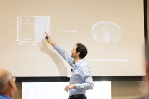
Assistant Professor Siniša Hrvatin studies states of stasis, such as mammalian torpor and hibernation, as a means to harness the potential of these biological adaptations to advance medicine. Photo: Steph Stevens
On April 19, 2023, the MIT School of Science hosted a breakfast talk featuring Siniša Hrvatin, an assistant professor of biology and a core member of the Whitehead Institute for Biomedical Research. Nergis Mavalvala, the Curtis (1963) and Kathleen Marble Professor of Astrophysics and dean of the School of Science, introduced Hrvatin to a group of alumni and guests gathered on the sixth floor of the Samberg Conference Center.
Hrvatin studies how animals initiate, regulate, and survive states of stasis. While Hrvatin’s research focuses on dormancy, Mavalvala urged the early-morning crowd to stay alert — which turned out to be an easy ask thanks to his lively talk titled “Biology of Mammalian Torpor and Hibernation.”
To survive extreme environments, many mammals from squirrels to bears have evolved to decrease their metabolic rate and body temperature, entering a state called torpor. Hibernation is marked by several consecutive periods of torpor. Hrvatin’s initial breakthroughs, though, were in mice models.
Aiming to locate the neurons responsible for initiating torpor, he observed the mice’s brain activity before, during, and after fasting-induced torpor, then mapped the activity onto a 3-D model of the brain. Eventually, his search narrowed to a region of neurons in the hypothalamus, including those responsible for metabolism and skin temperature.

Next, Hrvatin had to figure out if these neurons were actually responsible for initiating torpor in the mice. To do so, the researchers added receptors to neurons that were active during torpor, effectively adding an on/off switch. Hrvatin reflected that the idea that they could induce torpor by reactivating the neurons, at first, “seemed far-fetched.”
However, by screening more than 200 regions of the brain, the researchers were able to pinpoint a specific region of the hypothalamus, which they named avMLPA. When avMLPA neurons were stimulated in the mice, researchers observed a drop in their metabolic rate and core body temperature.
Using microfluidics, researchers proceeded to break down and sequence around 28,000 neurons in this region of the brain, identifying 36 different cell types. Only 342 neurons were active during torpor.
From there, the researchers leveraged neuroscience tools — installing fluorescent sensors of neuronal activity and optical fibers — to monitor the mice’s brains in real time. These tools demonstrated a connection so clear that researchers could predict when the mice were in torpor just by looking at the computer screen. What’s more, switching the same neurons off was shown to inhibit natural torpor in the mice.
“The results indicate that stimulating a population of neurons that are active during torpor, even in animals that aren’t actively fasting, is sufficient to induce this state,” confirmed Hrvatin.
Solving the many mysteries of torpor and hibernation, Hrvatin hopes, could lead to advancements in human medicine. Could these insights open the door for new treatments for metabolic disorders, or for muscle atrophy in bed-ridden patients? Might unlocking the mechanisms of how cells adapt to torpor improve organ transplants, during which organs are cooled down to minimize cell death, or even help to slow disease progression? What are the implications for slowing down epigenetic aging and potentially increasing the natural life span?
What if knowledge about states of suspended animation was applied to space travel?
“It is science fiction right now,” Hrvatin concluded. “But I really do believe that by studying the biology behind this, we can learn how to harness the potential in several medical fields.”
Sarah Costello | School of Science